Pulse Oximeter on the penis during penile implant surgery
Background:
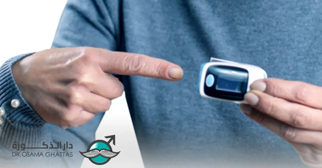
A pulse oximeter measures:
- Oxygen saturation (SpO₂) of blood in real time.
- Pulsatile flow, indirectly confirming perfusion.
- In penile implant surgery, the penis (especially the corpora cavernosa and glans) relies on good blood supply to maintain tissue viability. One theoretical use of a penile pulse oximeter would be to:
- Monitor real-time perfusion during surgery.
- Ensure no vascular compromise (e.g., from excessive traction, hematoma, or vessel injury).
Status of evidence?
- No standard practice:
There’s no current literature or guidelines suggesting that pulse oximetry is routinely used on the penis during implant surgery.
Closest analogs:
- Some research in reconstructive surgery (like phalloplasty) has explored monitoring penile flap perfusion using:
- Near-infrared spectroscopy (NIRS).
- Laser Doppler flowmetry.
- Occasionally, adapted pulse oximetry probes (e.g., on the glans or flap skin).
In penile prosthesis surgery, the main concerns are:
o Avoiding injury to the urethra, dorsal neurovascular bundle, and corpora.
o Preventing postoperative infection and erosion.
o Ensuring correct cylinder placement.
o Real-time perfusion monitoring isn’t seen as critical because:
• The implant sits inside the corpora cavernosa, which are well vascularized.
• Vascular injury would usually be apparent via bleeding or color change.
Potential Pros:
- Early detection of compromised perfusion:
If traction or device placement were to compress key vessels (dorsal artery), the pulse oximeter might detect a drop in perfusion before visible signs appear. - Innovation in high-risk cases:
For patients with vascular disease or previous surgeries, this might give extra safety reassurance. - Proof-of-concept for research:
Could be tested in research settings to validate whether penile SpO₂ changes correlate with complications.
Potential Cons & Challenges:
- Probe design:
Standard finger/toe pulse oximeters aren’t designed for penile anatomy, especially in flaccid or surgically manipulated tissue. Adapting a probe (e.g., ear clip or flexible wrap) could be tricky. - Movement artifacts:
Surgical manipulation of the penis creates significant movement, pressure changes, and traction, which would likely cause inaccurate or unstable readings. - Limited added value:
– Perfusion compromise during this surgery is rare and usually visible clinically.
– Major complications post-op (infection, erosion) is not directly tied to intraoperative perfusion in most cases.
– The penis receives its blood supply from three main arteries: the dorsal penile artery, the bulbourethral artery, and the cavernous artery. The glans penis is supplied primarily by the bulbourethral artery, while the corpora cavernosa are supplied by the cavernous arteries. Importantly, there is no direct vascular connection between these arterial systems. Therefore, even in the unlikely event that the cavernous artery is damaged during penile prosthesis surgery, the blood flow and oxygenation of the glans penis would remain unaffected. Furthermore, the surgical incision for prosthesis implantation is typically made on the ventral aspect of the penis or at the penoscrotal junction—well away from the dorsal neurovascular bundle, which contains the primary nerves and blood vessels of the penis. As a result, the risk of injuring these critical structures during this type of surgery is exceedingly low, especially compared to other penile procedures such as curvature correction or penile reconstruction, which require more extensive dissection near the neurovascular bundle. - Cost & complexity:
Adds extra steps and equipment to a procedure that is otherwise straightforward.
Conclusion:
- There’s no current evidence base or standard practice for using a pulse oximeter during penile implant surgery.
- The idea is theoretically interesting, especially for research, but faces practical challenges (probe fit, artifact, unclear clinical value).
- More sophisticated monitoring (like NIRS or laser Doppler) has seen limited use in complex penile reconstruction but not in prosthesis implantation.
Highlighted Reference:
Şenayli, Yeşim Andiran, Gülsen Keskin, Mine Akin, Atilla Şenayli, Rabia Ata, Gökhan Demirtaş, and Emrah Şenel. “A prospective study for an alternative probe site for pulse oximetry measurement in male patients with severe burn trauma: penile shaft.” Turkish Journal of Medical Sciences 53, no. 2 (2023): 504-510.
Background/aim
Authors widely use pulse oximetry in clinical monitoring of heart rate (HR) and peripheral oxygen saturation (SpO2) by attachment to the fingers; however, there can be a need for an alternative attachment site, especially for burned patients. We investigate the availability of a pulse oximeter probe attached to the penile shaft as an alternative site in pediatric male patients if all extremities became unavailable for pulse oximetry measurement due to severe burn and/or trauma.
Materials and methods
We designed a prospective comparative study in a training and research hospital. After local ethical committee approval, pediatric male cases eligible for penile and extremity pulse measurements were evaluated during general anesthesia for medical dressing and/or grafting due to severe burns. One probe was attached to the fingers of the unburned extremity, and the other was to the penile shaft. Furthermore, we recorded SpO2 and HR values at 5-min intervals; 0th (baseline), 5th, 10th and 15th minutes. We compared HR and SpO2 values measured by the finger probe with those measured by the penile probe.
Results
Data of 51 patients (median age, 2.9 years (interquartile range, 2.0-5.0 years)) in whom the duration of dressing was at least 15 min were analyzed. There was no significant difference either in comparisons of hemodynamic measurements (HR and SpO2) obtained by finger probe and by a penile probe for each measurement time. The Bland-Altman plot analysis reveals agreement for penile and finger probes with a mean bias value between 0.20 and 0.37 on HR and between 0.43 and-0.20 on SpO2.
Conclusion
This clinical trial demonstrated that pulse oximetry measurement under nonhypoxic conditions we could perform confidently using penile probes in pediatric male patients whose extremities are unavailable for measurement.
References:
- Nitzan, Meir, Amir Romem, and Robert Koppel. “Pulse Oximetry: Fundamentals and Technology Update.” Medical Devices (Auckland, N.Z.) 7 (2014): 231–39. https://doi.org/10.2147/MDER.S47319.
- McMorrow, Robert C., and Monty G. Mythen. “Pulse Oximetry.” Current Opinion in Critical Care 12, no. 3 (2006): 269–71. https://doi.org/10.1097/01.ccx.0000224873.16700.78.
- Petersen, Christine L., Tzu-Pin Chen, Joseph M. Ansermino, and Guy A. Dumont. “Design and Evaluation of a Low-Cost Smartphone Pulse Oximeter.” Sensors (Basel) 13, no. 12 (2013): 16882–93. https://doi.org/10.3390/s131216882.
- Jose, Binu, Rakesh Lodha, and Sushil K. Kabra. “Comparison of Two New Generation Pulse Oximeters with Arterial Oxygen Saturation in Critically Ill Children.” Indian Journal of Pediatrics 81, no. 12 (2014): 1297–1301. https://doi.org/10.1007/s12098-014-1381-z.
- Seifi, Shahram, Ali Khatony, Gholamreza Moradi, Ahmad Abdi, and Fariba Najafi. “Accuracy of Pulse Oximetry in Detection of Oxygen Saturation in Patients Admitted to the Intensive Care Unit of Heart Surgery: Comparison of Finger, Toe, Forehead and Earlobe Probes.” BMC Nursing 17, no. 15 (2018). https://doi.org/10.1186/s12912-018-0283-1.
- Clayton, D. G., R. K. Webb, A. C. Ralston, D. Duthie, and W. B. Runciman. “Pulse Oximeter Probes: A Comparison Between Finger, Nose, Ear, and Forehead Probes under Conditions of Poor Perfusion.” Anaesthesia 46, no. 4 (1991): 260–65. https://doi.org/10.1111/j.1365-2044.1991.tb11492.x.
- Morey, Timothy E., Michael J. Rice, Thomas Vasilopoulos, David M. Dennis, and Richard J. Melker. “Feasibility and Accuracy of Nasal Alar Pulse Oximetry.” British Journal of Anaesthesia 112, no. 6 (2014): 1109–14. https://doi.org/10.1093/bja/aeu095.
- Robertson, R. E., and R. F. Kaplan. “Another Site for the Pulse Oximeter Probe.” Anesthesiology 74, no. 1 (1991): 198. https://doi.org/10.1097/00000542-199101000-00036.